Knee Muscle Anatomy Mri - Common Mistakes And Pitfalls In Magnetic Resonance Imaging Of The Knee. Mri for evaluating knee pain in older patients: Magnetic resonance imaging (mri) tests involve large machines that use radio wave energy pulses and a magnetic field to produce images of the shoulder (2). When interpreting the proton density images it. Plantaris acts weakly to plantar flex the foot and flex the knee. Mri knee anatomy scroll using the mouse wheel or the arrows.
Use the checklist to quiz yourself. Weak adductor muscles may cause knee instability and adductor strain (2). Anatomical structures of the lower limb (hip, thigh, knee, leg, ankle and foot) and specific regions (compartment of the lower. 12 photos of the knee muscle anatomy mri. Anatomy basic knee mri checklist.
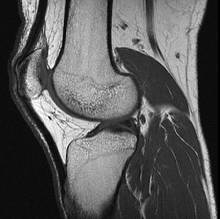
Accessory Muscles Of The Knee Radsource from radsource.us The main knee muscles are the quadriceps, hamstrings and calf muscles. This mri knee sagittal cross sectional anatomy tool is. From superficial to deep includes the pes anserinus tendons, semimembranosus tendon, tibial collateral ligament, meniscofemoral and meniscotibial ligaments, and the medial meniscus. Naturally, in order to assess pathologic knee imaging, it is necessary to know the appearance of a normal knee mri. Magnetic resonance imaging is particularly well suited for the medical evaluation of the musculoskeletal (msk) system including the knee, shoulder, ankle, wrist and elbow. Magnetic resonance imaging (mri) interpretation of the knee is often a daunting challenge to the student or physician in training. Knee muscles need to have both good strength and flexibility. This article is based on a presentation given by david rubin and adapted for the radiology assistant by robin smithuis.
To realign the anterior cruciate ligament parallel with the sagittal imaging plane.
They are attached to the femur (thighbone), tibia (shinbone), and fibula (calf bone) by fibrous tissues called ligaments. These motions of the knee allow the body to perform such important movements as walking, running, kicking, and jumping. Thigh muscles also protect neurovascular structures as they go through the proximal hip joint to the knee and lower leg (3). Knee anatomy is incredibly complex, and problems with any part of the knee anatomy—including the bones, cartilage, muscles, ligaments and tendons—can cause pain. Naturally, in order to assess pathologic knee imaging, it is necessary to know the appearance of a normal knee mri. In this presentation mri anatomy biceps femoris muscle. Anatomical structures of the lower limb (hip, thigh, knee, leg, ankle and foot) and specific regions (compartment of the lower. Knee mri anatomy of the knee anterior cruciate ligament pet ct biceps study health fitness radiology. This article is based on a presentation given by david rubin and adapted for the radiology assistant by robin smithuis. Abnormal anatomy with normal signal, i.e. These muscles work in groups to flex extend and stabilize the knee joint. Magnetic resonance imaging (mri scan): Serves as a paid consultant to or is an employee of conformis inc.;
Injuries such as anterior cruciate ligament, meniscus and rotator cuff tears are all easily diagnosed when there is a firm understanding and knowledge of human anatomy. Anatomy of the knee bones around the knee. Tibial tuberosity with distal patella tendon insertion. Magnetic resonance imaging is particularly well suited for the medical evaluation of the musculoskeletal (msk) system including the knee, shoulder, ankle, wrist and elbow. Anatomy basic knee mri checklist.
Knee Anatomy Mri Knee Coronal Anatomy Free Cross Sectional Anatomy from mrimaster.com Prescribe sagittal plane off axial images with line parallel to bony glenoid. Can also generate proton density images. The images may also help physicians to distinguish normal, healthy tissues from dead tissues(2). Mri for evaluating knee pain in older patients: They are attached to the femur (thighbone), tibia (shinbone), and fibula (calf bone) by fibrous tissues called ligaments. These muscles work in groups to flex extend and stabilize the knee joint. These motions of the knee allow the body to perform such important movements as walking, running, kicking, and jumping. The muscles of the knee include the quadriceps, hamstrings, and the muscles of the calf.
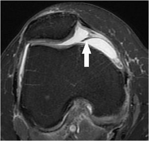
Knee muscle anatomy axial mri :
Magnetic resonance imaging (mri) interpretation of the knee is often a daunting challenge to the student or physician in training. These muscles work in groups to flex extend and stabilize the knee joint. This mri knee sagittal cross sectional anatomy tool is absolutely free to use. Plantaris can have variable size, but in most cases is difficult to demonstrate on routine mri studies. It is considered a vestigial muscle, and can be used as a tendon graft in reconstructive orthopedic surgery. By now you probably know that the anatomy is deceptively complex, combinations of injuries can be challenging, and of course the referring clinician's. The common peroneal nerve typically courses downward within abundant fat posterior to the short head of the biceps femoris muscle and superficial to the lateral head of the gastrocnemius muscle, but. Atlas of knee mri anatomy. 4, infrapatellar fat pad of hoffa. Louis, usa and the rijnland hospital in leiderdorp, the netherlands. General anatomy and musculoskeletal system. Anterior and posterior cruciate ligaments. In conclusion, we describe the normal mri anatomy of the distal biceps femoris and the relationship of this muscle with the common peroneal nerve.
These muscles work in groups to flex extend and stabilize the knee joint. Can also generate proton density images. Anatomical structures of the lower limb (hip, thigh, knee, leg, ankle and foot) and specific regions (compartment of the lower. Medical images from an mri allow medical professionals to distinguish body tissues, including the meniscus (shock absorbers in the knee), cartilage, tendons, and ligaments. The common peroneal nerve typically courses downward within abundant fat posterior to the short head of the biceps femoris muscle and superficial to the lateral head of the gastrocnemius muscle, but.
Knee Mri Radiology Key from radiologykey.com Naturally, in order to assess pathologic knee imaging, it is necessary to know the appearance of a normal knee mri. Knee muscles need to have both good strength and flexibility. Mri for evaluating knee pain in older patients: General anatomy and musculoskeletal system. Thigh muscles also protect neurovascular structures as they go through the proximal hip joint to the knee and lower leg (3). Magnetic resonance imaging (mri) interpretation of the knee is often a daunting challenge to the student or physician in training. Use the checklist to quiz yourself. Anatomical structures of the lower limb (hip, thigh, knee, leg, ankle and foot) and specific regions (compartment of the lower.
Can also generate proton density images.
Naturally, in order to assess pathologic knee imaging, it is necessary to know the appearance of a normal knee mri. These muscles work in groups to flex extend and stabilize the knee joint. Coronal anatomy of the knee. These muscles work in groups to flex, extend and stabilize the knee joint. Atlas of knee mri anatomy. Mri wrist anatomy scroll using the mouse wheel or the arrows. This article is based on a presentation given by david rubin and adapted for the radiology assistant by robin smithuis. Anterior and posterior cruciate ligaments. 4, infrapatellar fat pad of hoffa. Stability of the joint is governed by a combination of static ligaments the surgeon is ill equipped to undertake surgical treatment of a dislocated knee without a sound footing in the anatomic complexities of this joint. The knee joint is the junction of the thigh and leg. Richolt j.a., jakab m., kikinis r. Doctors may recommend a knee mri if a patient experiences the following(3):